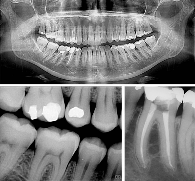
Extraordinary X-ray Image Quality
CADI® uses powerful tools commonly found in medical imaging to bring optimal results with any digital equipment. CADI® keeps all the raw data from the x-ray sensor and uses a powerful 16-bit viewer and the unique Windowing tool permits the user to interpret different grayscale representations “Windows” of this data. Eliminate the parts of the image that don’t contribute to diagnostics. This allows CADI® to offer extraordinary image quality with a variety of brands of sensors and phosphor plate systems.
Actual CADI images
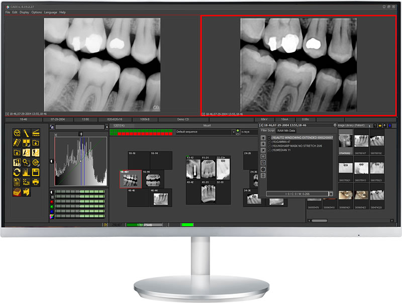
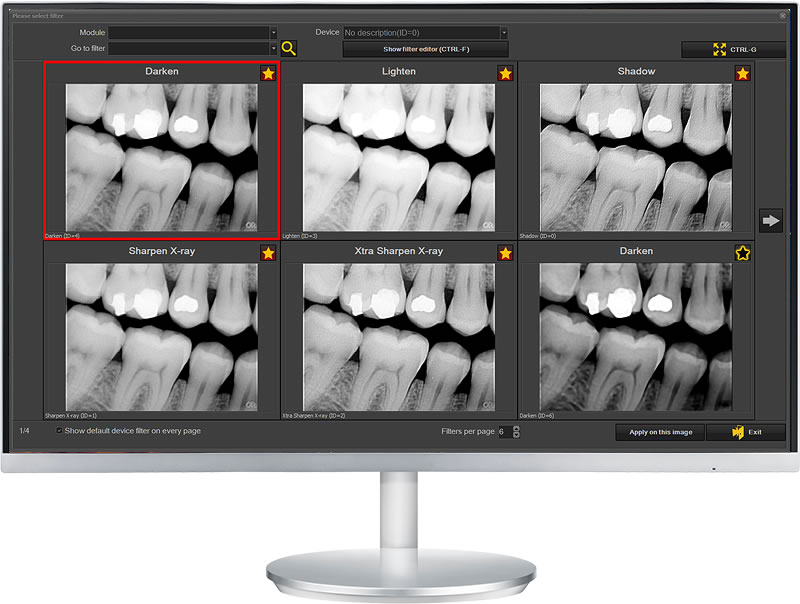
Real Time Filtering and Filter Window
Use pre-set or customizable x-ray real-time filters to visualize the most diagnostic images possible. The raw image data is always available, so you can unclick a filter at any time. Click on the “Filter” window and simultaneously zoom to view 6 different filtered images.
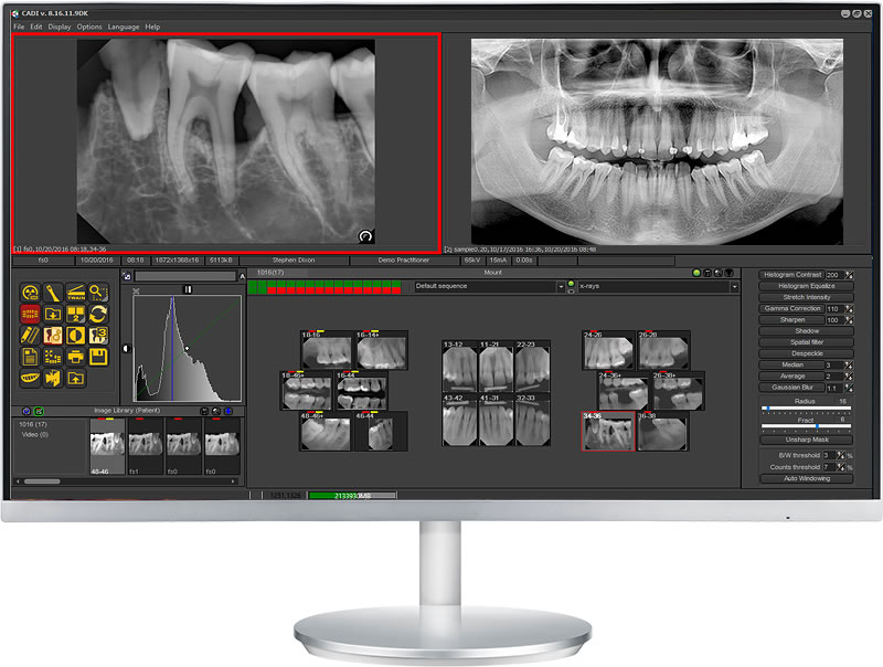
Powerful Status-mount Configuration
That include exam selection and tooth history views
From a single status-mount (preconfigured or customizable), you can select a preconfigured intra-oral x-ray exam (number of teeth, tooth numbers, orientation and order). Images are all stored in a single multi-depth mount. View the current exam, x-rays of any tooth, and history of x-rays on that tooth.
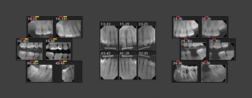
X-ray mount
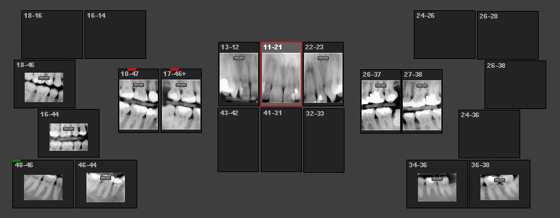
Vertical bitewing x-ray mount
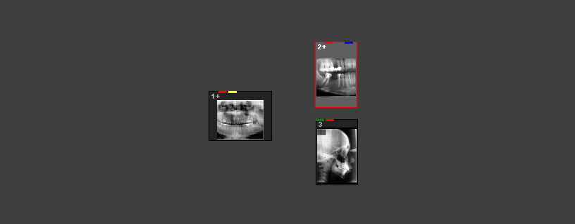
Pan & Ceph mount
Intra-oral camera mount
Digital camera mount
Ortho mount
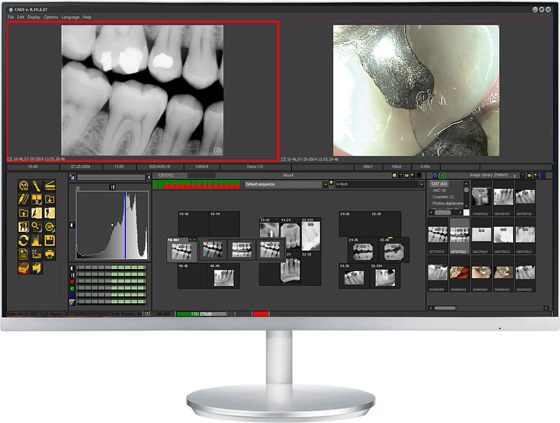
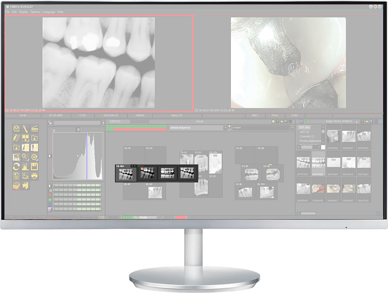
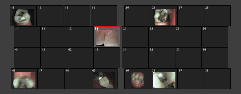
Intra-oral Camera
Acquire, store, and view intra-oral camera images by tooth number
Click on the tooth in the status-mount and images are tagged and stored in the tooth in the mount. Use a CADI-customized intra-oral camera and move through the mount to take intra-oral camera images.
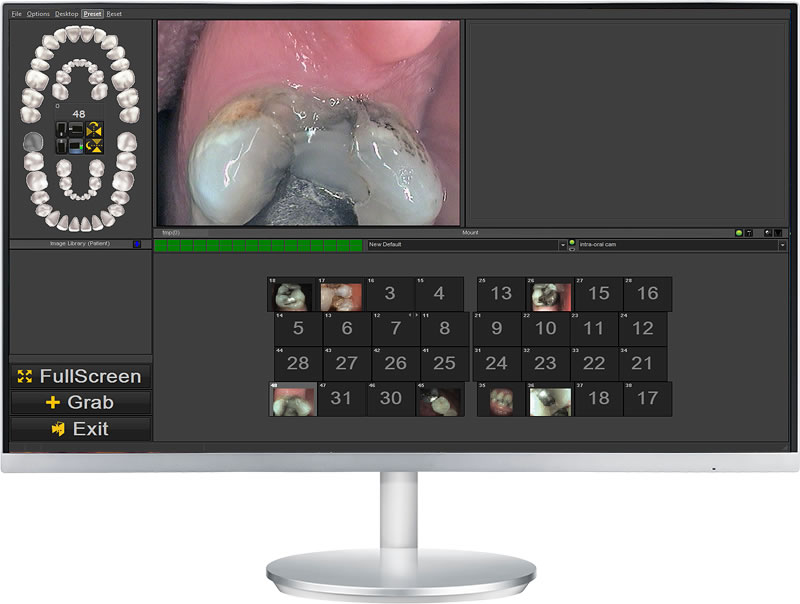
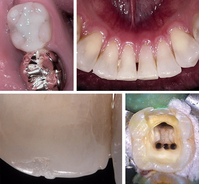
Actual CADI images
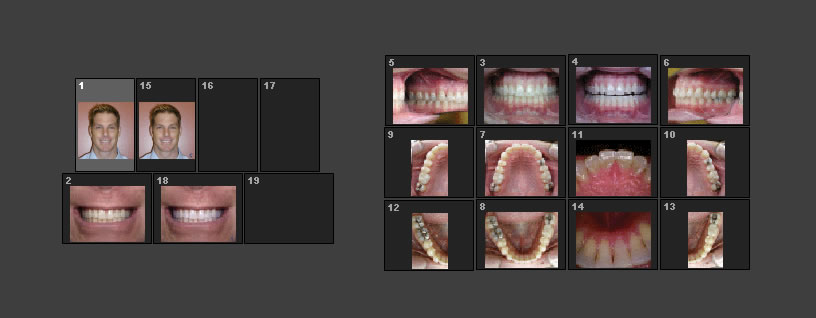
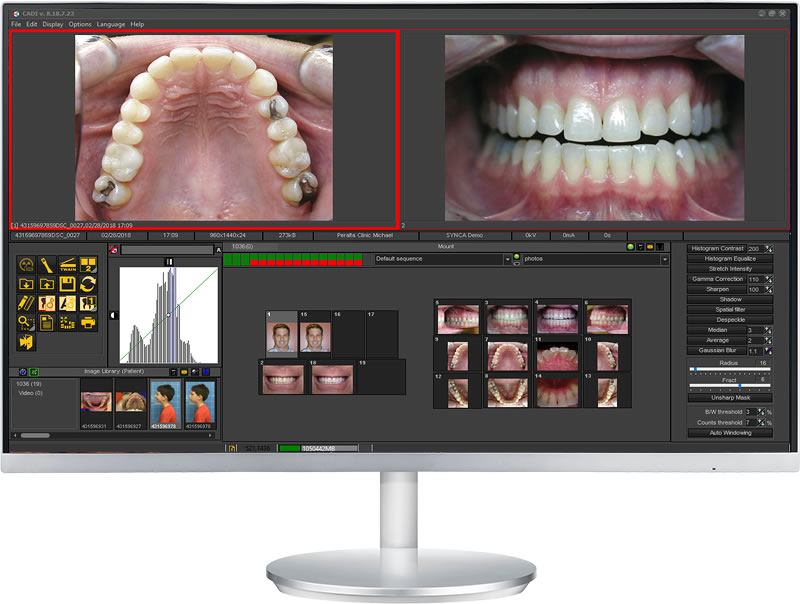
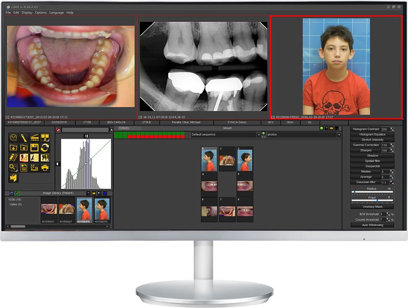
Digital Camera
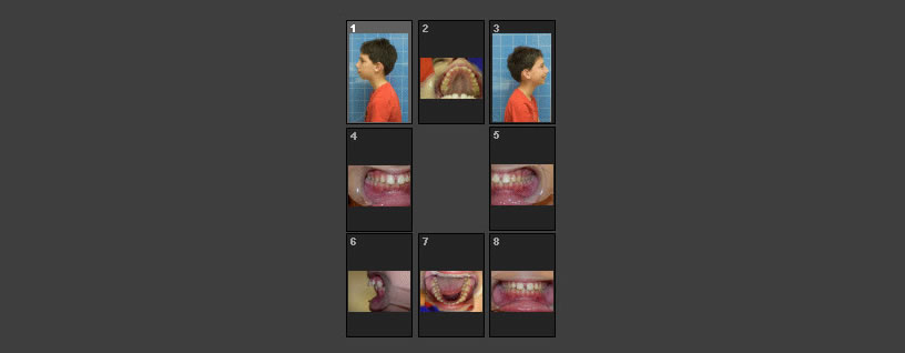
Set up your new patient exam, or use the AAO status mount for orthodontic cases and automatically import digital camera images for easy storage and viewing.
Actual CADI images
Actual CADI images
3D Viewer and 3D launcher
From the CADI® patient main screen, you can launch your 3D imaging software. Use the inexpensive CADI® 3D Viewer to view images on different workstations.
Open 3D images in CADI®. The 3D Volume viewer provides 4 different volume construction techniques: MPR, VRT, MIP and SSD. With the 2D stack overview, navigate through the images or different slices. Reconstruct a panoramic image by using the MPR/panoramic polygon feature.
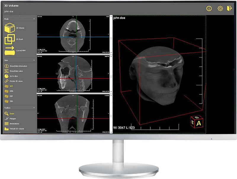
3D volume viewer
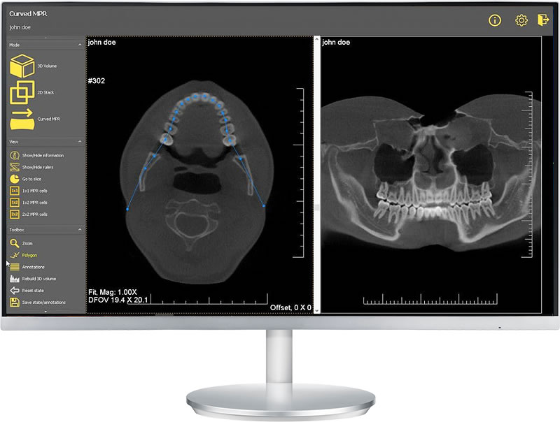
Curved MPR